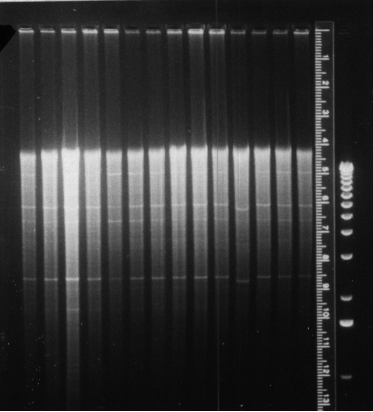

The figure on the left shows a photograph of a 0.7% agarose gel that has 14 different samples loaded on it (plus molecular weight marker in the far right lane and a glowing ruler used for analysis of the results). The samples are genomic DNA isolated from different strains of the unicellular green alga Chlamydomonas reinhardtii. Each sample of DNA has been digested with the same restriction enzyme (EcoRI). Notice that the DNA does not appear as a series of discrete bands but rather as a smear. [Why is that?]
This DNA was transferred to nitrocellulose and then probed with a radioactive fragment of DNA that is also present in a subset of the DNA isolated from the different strains. The figure on the right is a copy of the X-ray film and reveals which strains contain the target DNA and which ones do not.
Return to the Biology Course Materials
Register in Our Guestbook and Submit Comments
© Copyright 2005 Department of Biology, Davidson
College, Davidson, NC 28036
Send comments, questions, and suggestions to: macampbell@davidson.edu