Monoclonal antibodies (MAbs for short) are an essential part of a molecular biologist's toolkit. But what exactly are these molecules? How and why are they used so extensively in molecular biology? The purpose of this web page is to answer these questions as best it can, and to direct you to further resources on the subject should you wish to research the topic more thoroughly.
Antibodies are proteins produced by the B lymphocytes of the immune system in response to foreign proteins, called antigens. Antibodies function as markers, binding to the antigen so that the antigen molecules can be recognized and destroyed by phagocytes. The part of the antigen that the antibody binds to is called the epitope. The epitope is thus a short amino acid sequence that the antibody is able to recognize (Campbell NA, 1996).
Two features of the antibody-epitope relationship are key to the use of monoclonal antibodies as a molecular tool.
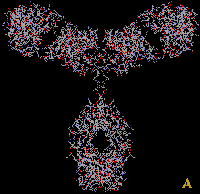
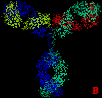
Structurally antibodies are proteins consisting of four polypeptide chains. These four chains form a quaternary structure somewhat resembling a Y shape. Figure 1 shows the three dimensional structure of immunoglobulin G, a typical antibody, and its schematic representation.
Figure 1. A -- 3-D representation of immunoglobulin G; B -- 3-D representation of immunoglobulin G color labelled to show the four polypeptide chains. These 3-D pictures are derived from the Rasmol representation of immunoglobulin G on the Davidson College Biology department web server.
Each B cell in an organism synthesizes only one kind of antibody. In an organism, there is an entire population of different types of B cells and their respective antibodies that were produced in response to the various antigens that the organism had been exposed to. However to be useful as a tool, molecular biologists need substantial amounts of a single antibody (and that antibody alone). Therefore we need a method to culture a population of B cells derived from a single ancestral B cell, so that this population of B cells would allow us to harvest a single kind of antibody. This population of cells would be correctly described as monoclonal, and the antibodies produced by this population of B cells are called monoclonal antibodies. In contrast, antibodies obtained from the blood of an immunized animal are called polyclonal antibodies.
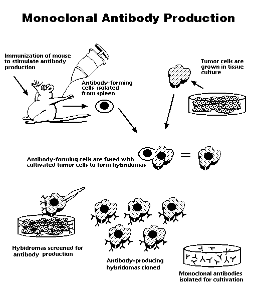
The production of monoclonal antibodies was pioneered by Georges Kohler and Cesar Milstein in 1975. Let us see how their method, now tried and tested for over 20 years, would be applied in a particular case.
In order for us to isolate a B lymphocyte producing a certain antibody, we first have to induce the production of such a B cell in an organism. For example, if we need an antibody for avian SERCA2 protein, we would inject the protein into a mouse. This is typically done in two doses, an initial "priming" dose and a second "booster" dose 10 days later (Campbell MA, pers. comm.). Since the protein is of foreign origin, the mouse immune system recognizes it as such and soon some of the B cells in the mouse would begin production of the antibody to avian SERCA2.
A sample of B cells is extracted from the spleen of the mouse and added to a culture of myeloma cells (cancer cells). The intended result is the formation of hybridomas, cells formed by the fusion of a B cell and a myeloma cell. The fusion is done by using polyethylene glycol, a virus or by electroporation (Campbell MA, pers. comm.)
The next step is to selct for the hybridomas. The myeloma cells are HGPRT- and the B cells are HGPRT+. HGPRT is hypoxanthine-guanine phosphoribosyl transferase, an enzyme involved in the synthesis of nucleotides from hypoxanthine, an amino acid (Biotech Resources,1995-7). The culture is grown in HAT (hypoxanthine-aminopterin-thymine) medium, which can sustain only HGPRT+ cells (Biotech Resources, 1995-7). The myeloma cells that fuse with another myeloma cell or do not fuse at all die in the HAT medium since they are HGPRT-. The B cells that fuse with another B cell or do not fuse at all die because they do not have the capacity to divide indefinitely. Only hybridomas between B cells and myeloma cells survive, being both HGPRT+ and cancerous.
The initial collection of B cells used is heterogenous, i.e. they do not all produce the same antibody. Therefore the hybridoma population too does not produce a single antibody. There is also another complication. A hybridoma cell is initially tetraploid, having been formed by the fusion of two diploid cells. However the extra chromosomes are somehow lost in subsequent divisions in a random manner (Campbell MA, pers. comm.). This means that we cannot be certain that the hybridomas will all produce the desired antibody or even any antibody at all. Screening is required to decide which hybridoma cells are actually producing the desired antibody.
Each hybridoma is cultured and screened after doing SDS-PAGE (sodium dodecyl sulfate - polyacrylamide gel electrophoresis) and Western blots. The probe used is the epitope of the antibody that is desired, which may be labeled by radioactivity or immunofluorescence. Once we are sure that a certain hybridoma is producing the right antibody, we can culture that hybridoma indefinitely and harvest monoclonal antibodies from it.
Figure 2 summarizes the procedure for the production of monoclonal antibodies.
Figure 2. A summary of the process of monoclonal antibody production. (Source: Biotech, 1989)
Monoclonal bodies have a variety of academic, medical and commercial uses. It would be impossible to list all of these here. But the following list should indicate how ubiquitous monoclonal antibody technology has become in biotechnology.
The following web sites have more information on monoclonal antibodies, as well as on antibodies in general.
Biotech Resources. 1989. Monoclonal antibody technology -- the basics.
<http://www.gene.com/ae/AB/IE/Monoclonal_Antibody.html>:
Accessed 1998 18 Feb.
Biotech Resources. 1989. Monoclonal antibody production. <http://www.gene.com/ae/AB/GG/monoclonal.html>:
Accessed 1998 18Feb.
Biotech Resources. 1995-7. Life ScienceDictionary. <http://biotech.chem.indiana.edu/search/dict-search.html>:
Accessed 1998 17 Feb.
Campbell, N.A. 1996. Biology - 4th Ed. The Benjamin/Cummings Publishing
Co., CA, pp. 862-869.
Chaudhuri, T.R., Zinn, K.R., Morris, J.S.,
McDonald, G.A., Llorens, A.S., and Chaudhuri, T.K. 1994. Human
monoclonal antibody developed against ovarian cancer cell surface antigen.
<http://www.thriveonline.com/@@EaXTTgUA2XVOywjU/thrive/health/Library/CAD/abstract8127.html>:
Accessed 1998 18 Feb.
Fratella, J. 1998. Research Probes Custom Made in Monoclonal Antibody
Facility. <http://www.orst.edu/Dept/research/asti/public_html/connect/con72/probe.htm>:
Accessed 1998 18 Feb.
Orrs, A. 1997. FDA approves monoclonal antibody for cancer treatment.
<http://www.nando.com/newsroom/ntn/health/112797/health7_7347_noframes.html>:
Accessed 1998 18 Feb..
P/S/L Consulting Group, Inc. 1997. Combination HIV-IG/monoclonal antibody
dose neutralizes AIDS virus. <http://www.pslgroup.com/dg/196b2.htm>:
Accessed 1998 18 Feb.
Transweb. 1996. Monoclonal antibodies and OKT3. <http://www.transweb.org/drugs/okt3.html>:
Accessed 1998 18 Feb.
Wang SP, Holmes KK, Knapp JS, Ott S, Kyzer. Immunologic classification
of Neisseria gonorrhoeae with immunofluorescence. J Immunol 1977;119:794-803.