
*This website was produced as an assignment for an undergraduate course at Davidson College.*
Human Immunodeficiency Virus Glycoprotein 120
| Figure 1. Ribbon diagram of HIV gp120 in complex with CD4. The gp120 is shown in red; CD4 is shown in yellow. The gp120 facilitates viral entry into human host cell. The protein binds to CD4, causing a conformation change in the envelope, allowing entry. Of particular note is the phenylalanine residue reaching into the recesses of gp120. (Kwong, 1998. Permission granted from the author). |
Introduction:
The AIDS pandemic is arguably the most critical health crisis facing the world today. The eradication of the Human Immunodeficiency Virus (HIV) has been a top priority for President Bush, Bono, and many others. On February 13, 2005, newspapers broke with a story describing a recently infected individual in New York City who showed little response to any drug therapy (Click here to go to article—registration required). What was more alarming was that his condition has degenerated to immune failure during a period of about two months. While some scientists are pointing to an already weakened immune system as a possible reason for the rapid onset, the emergence of this new strain points to one of the main defenses viruses have—they can rapidly mutate to stay one step ahead of the immune system (Santora and Altman, 2005). However, there are highly conserved portions of the HIV that are necessary for the virus to infect cells. The protein gp120 lies on the outer envelope of the HIV, binds to a receptor on a human cell, and mediates entry of the virus. The amino acid residues responsible for this binding are highly conserved among various strains (Kwong, 1998). |
Function of gp120:
| In order to understand the structure of gp120, it is perhaps best to understand the function of gp120 first. The exact mechanism by which the virus enters the cell is unknown; however, it is known that gp120 plays a critical role. The protein's role is threefold: to seek receptors suitable for viral entry, to fix the viral particle to the cell, and to assist and direct the injection of viral material. The envelope protein primarily functions to bind to the protein CCR5. This binding induces a change in the confirmation of gp120; it is through this binding complex that the three dimensional structure of the protein itself was originally discovered. The protein is covered with a number of glycosylation sites. These sites have oligosaccharides attached to them, which prevents detection by the immune system. Another evasion technique employed by the protein is that critical binding sites on the protein are found recessed in the surface, allowing the variable regions of the protein to sterically hinder antibodies (Kwong, 1998). |
Structure of gp120:
| The structure of gp120 has "no precedent," according to those who deduced its structure (Kwong, 1998). The inner domain contains no homologous sequences to any other organism, while there is some sporadic homology among sections of the outer domain. One outer section bears homology to a dehydrase promoter, while another section has some sequence homology to a dUTP pyrophosphatase. dUTP pyrophosphatase is an enzyme found in viruses related to HIV, although there is no evidence yet that links this enzyme to the protein coat. The protein itself shows wide variability in sequence among strains of the virus, but there are some sequences that show some conservation. In general, the inner domain is much more conserved than the outer domain, most likely indicating some critical function. The glycosylation sites themselves are also highly conserved (Kwong, 1998). |
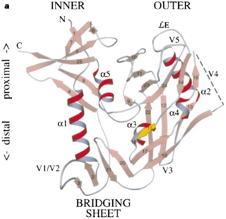
| Figure 2. Structure of gp120. In relation to previous figure, this structure has been rotated 180 degrees. Note the presence of outer and inner domains, and the cavity located between them. The CD4/gp120 binding site is inside this cavity. (Kwong, 1998. Permission granted from the author). |
CD4 and gp120:
| Part of the CD4 protein wedges itself directly in between the outer and inner domains of gp120. This cavity in between the domains is remarkably well conserved between different strains, and contains no glycosylation sites. The attraction between CD4 and gp120 is mostly electrostatic, with the primary end of CD4 attracted to the primarily negative cavity of gp120. There are also 219 van der Waals attractions and 12 hydrogen bonds formed between the two proteins. It is interesting to note that nearly 23% of intermolecular attractions come from the Phenylalanine residue #43 on CD4, and 57% of all gp120 interactions with CD4 stem from two separate amino acid sequences of about 5 residues each. Apart from these interactions, however, the region where gp120 binds to CD4 is highly variable. The cavity formed where CD4 binds to gp120 is much larger than the region of CD4 that finds itself in the region. Because this cavity does not seem specific for CD4, the authors speculate that the cavity is created by a conformational change in the gp120 protein when CD4 binds. |
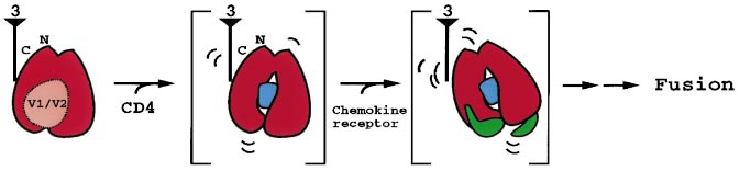
| Figure 3. Proposed confirmation change in gp120 following binding with CD4. Note that the red protein represents gp120, and the blue circle represents the larger cavity created by the conformational shift following binding with CD4. (Kwong, 1998. Permission granted from the author). |
References:
Kwong PD, Wyatt R, Robinson J, Sweet RW, Sodroski J, Hendrickson WA. 1998. Structure of an HIV gp120 envelope glycoprotein in complex with the CD4 receptor and a neutralizing antibody. Nature 393: 648-659. Santora, M and Altman, L. 2005 Feb. 12. Rare and Aggressive H.I.V. Reported in New York. New York Times. <http://www.nytimes.com/2005/02/12/health/12aids.html> Accessed 15 Feb. 2005. |
Questions? Comments? Concerns? Email me: wigreendyke@davidson.edu