In multicellular organisms, all the cells are identical in their DNA but the proteins vary tremendously. Therefore, it would be very useful if we could separate cells that are phenotypically different from each other. In addition, it would be great to know how many cells expressed proteins of interest, and how much of this protein they expressed. Fluorescence Activated Cell Sorting (FACS) is a method that can accomplish all these goals.
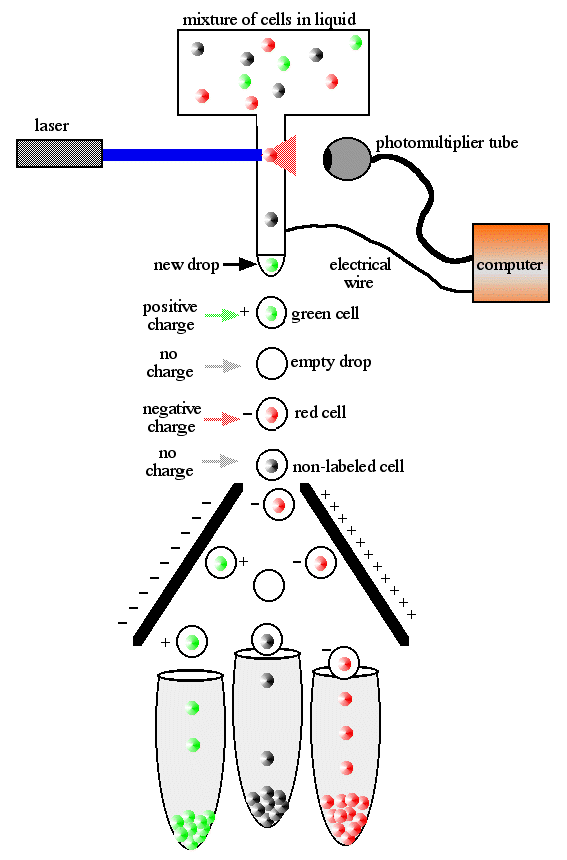
The process begins by placing the cells into a flask and forcing the cells to enter a small nozzle one at a time (figure 1). The cells travel down the nozzle which is vibrated at an optimal frequency to produce drops at fixed distance from the nozzle. As the cells flow down the stream of liquid, they are scanned by a laser (blue light in figure 1). Some of the laser light is scattered (red cone emanating from the red cell) by the cells and this is used to count the cells. This scattered light can also be used to measure the size of the cells.
If you wanted to separate a subpopulation of cells, you could do so by tagging those of interest with an antibody linked to a fluorescent dye. The antibody is bound to a protein that is uniquely expressed in the cells you want to separate. The laser light excites the dye which emits a color of light that is detected by the photomultiplier tube, or light detector. By collecting the information from the light (scatter and fluorescence) a computer can determine which cells are to be separated and collected.
Figure 1. Diagram of FACS machine. Cells have been fluorescently tagged with either red or green antibodies, though not every cell expresses the epitope and therefore some are not tagged either color.
The final step is sorting the cells which is accomplished by electrical charge. The computer determines how the cells will be sorted before the drop forms at the end of the stream. As the drop forms, an electrical charge is applied to the stream and the newly formed drop will form with a charge. This charged drop is then deflected left or right by charged electrodes and into waiting sample tubes. Drops that contain no cells are sent into the waste tube. The end result is three tubes with pure subpopulations of cells. The number of cells is each tube is known and the level of fluorescence is also recorded for each cell.
FACS data collected by the computer can be displayed in two different ways. What we want to know is how many cells of each color were sorted. In the first example (figure 2), we see the intensity of the green or red fluorescence is plotted on the X-axis and the number of cells with each level of flourescence is plotted on the Y-axis. In this example, there were twice as many red cells sorted as green or unlabeled cells, but the level of light was greater from the green cells than the red cells. This method is best if all cells are either green, red or unlabeled and no cells are labeled both colors.
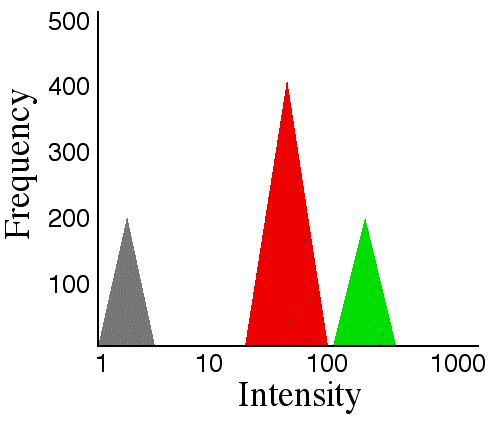
Figure 2. Quantifying FACS data. This graph shows the number of cells (Y-axis) and the level of fluorescence emitted (X-axis) by the labeled cells. Many different colors can be plotted on this graph, but cells should not be labeled by more than one color.
In figure 3, we see a different way to display the same data. The X-axis plots the intensity of green fluorescence while the Y-axis plots the intensity of red fluorescence. The individual black dots represent individual cells and we are not supposed to count the dots but just look at the relative density of dots in each quadrant. From this graph, we can see there were no cells labeled both red and green (top right) and many cells that were unlabeled (bottom left). The number of green-labeled cells (bottom right) is about the same as the number of unlabeled cells, but the number of red-labeled cells (top left) is about twice that of the other two categories of cells. Again, we can see that the level of fluorescence was higher in the green cells than the red ones. This method of graphing the data is especially useful if cells are present that have been labeled both red and green.
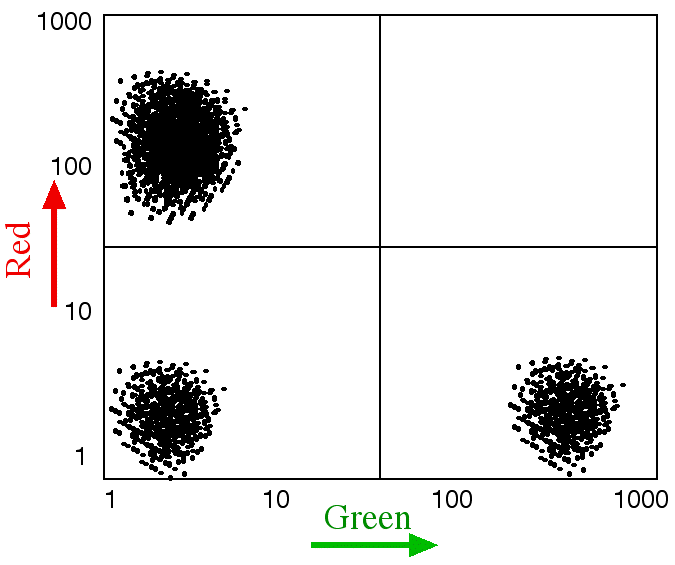
Figure 3. Quantification of FACS data. This graph compares the number of cells labeled by two colors - red (Y-axis) and green (X-axis). The intensity of the emitted light increases as indicated by the arrows. The number of cells at each intensity is shown by the number of dots where each dot represents a single cell. This graph does not work for more than two colors but it works well when individual cells can be labeled by both colors at the same time.
![]()
![]()
© Copyright 2001 Department of Biology, Davidson College, Davidson, NC 28036
Send comments, questions, and suggestions to: macampbell@davidson.edu