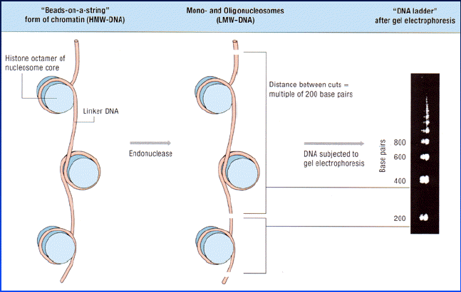
Here are some images that help you understand how cells undergoing programed death can have their DNA cut into 200 bp fragments. The images were produced by Roche Molecular Biochemicals at <http://biochem.roche.com/apoptosis/slide1.htm> and can be viewed as a part of a very good slide show.
Figure 1. This is how DNA is wound around histone complexes inside the nuclei of your cells and how the chromosomes are cut into 200 bp fragments in apoptotic cells. Images used with permission from Roche Molecular Biochemicals at <http://biochem.roche.com/apoptosis/slide1.htm>
Figure 2. If the genomic DNA of apoptotic cells were run on a gel, you would see the pattern in the right lane. Note the regularly spaced bands of DNA, each 200 base pairs different in length.The left lane shows genomic DNA isolated from healthy cells. Images used with permission from Roche Molecular Biochemicals at <http://biochem.roche.com/apoptosis/slide1.htm>
For a more information about the complex pathways leading to apoptosis, try out another great web page from Roche.
Return To Immunology Main Page
Return to Immunology Reading Schedule
© Copyright 2002 Department of Biology, Davidson
College, Davidson, NC 28036
Send comments, questions, and suggestions to: macampbell@davidson.edu