
This web page was produced as an assignment for an undergraduate course at Davidson College.
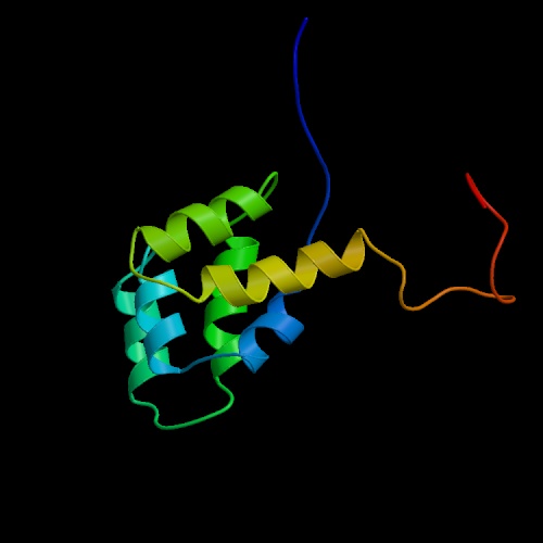
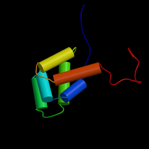
Figures 1 and 2: These images show ribbon and cylinder representations of the structure of the Fas death domain. The death domain consists of six antiparallel a helices. Death domains have a tendency to self-associate and are found in many proteins involved with apoptosis, including Fas, TNFR and FADD. The structure was solved using NMR-spectroscopy by Huang, Eberstadt, Olejniczak, Meadows, and Fesik. The images are public property as a part of the international Protein Data Bank.