This web page was produced as an assignment for an undergraduate course at Davidson College
Evolution teaches that all species emerged from a common ancestor. Some ancestors, such as those of humans and primates, are relatively close in relation, while others, such as the common ancestor of humans and yeast, are relatively far apart in relation. Since all species came from a common ancestor, the genomic and proteomic codes must also have been the same at one point in time. Many species still share similar protein codes as a result of evolving from a common ancestor and as a result of conservation of vital codes within the genome. Similar coding sequences between species indicate may indicate common ancestry and sequence conservation. Studying orthologs, or genes that are in different species but have evolved from one another, sheds light on the importance of conservation within the protein sequence and the relative amount of evolution between species.
Model organisms are often used for ortholog searches, but any species with a sequenced proteome will do. To search for phenylalanine hydroxylase (PheOH) orthologs, I used the protein sequence found here from NCBI. I performed a Blast search using the protein code, and found very similar orthologs in many species: Mus musculus (mouse), Rattus norvegicus (rat), Gallus gallus (chicken), Danio rerio (zebrafish), Canis familiaris (dog), and Pongo pygmaeus (orangutan). (Find a more complete list of species with percent identities, positives, and e-values here.)
It is worthy to note where in the protein sequence the orthologs were similar. In the first assignment, Kobe and colleagues discuss the importance of the area around amino acids 111-117 as a possible site of hinging, where the protein could change shape to cleave phenylalanine. Because this site is thought to perform an important function of the protein, it stands to reason that this would be a an area that is highly conserved throughout species. This is not the case. Amino acids 111-117 are not as conserved as areas suggesting that this sequence is not as functionally important as believed by Kobe et al. Instead, the differences in the sequences show an increasing distance from a common ancestor.
Homo sapien aa111-117: |
RDKKKDT |
Different from Human |
| Pongo pygmaeus aa106-112: | RDKKKDT |
RDKKKDT |
Mus musculus aa111-117: |
RNKQKDT |
RDK+K+T |
Rattus norvegicus aa111-117: |
RTKKKDT |
RDK+K+T |
| Danio rerio aa109-115: | RDKEKNT |
R+K+KDT |
| Gallus gallus aa105-111: | RDKEKNT |
R-KKKDT |
| Drosophila melanogaster aa108-114: | RDYKDNA |
RD-K - + - |
| Aedes aegypti (mosquito)aa105-113: | RDYKDNEAT |
RD-K-+(EA)T |
| Geodia cydonium (sponge) aa110-116: | RDPGEDE |
RD- -+D- |
| Branchiostoma floridae (lancelet) aa96-101: | TPPTEDS |
- - - -+d+ |
Table 1: Human PheOH amino acids 111-117 are listed at the top in red. Species are identified to the left with the amino acids lining up to the human sequence. The far right column shows the differences from the human sequence; letters indicate a match to the human reference, + indicates a similar amino acid, - indicates a dissimilar amino acid, and letter in ( ) indicate amino acids added into the species' sequence. Sequences and comparisons are from Blast.
Most of the orthologs, while differing at various points throughout the sequence, show an increased amount of similarity from approximately amino acid 221 to 360 (from my observations.) A second Blast search of just this subset of the sequence reveals a 98% identical and 100% positive match to Mus musculus, Rattus norvegicus, and Canis familiaris, 92% identical and 97% positive match to Gallus gallus, 88% identical and 95% positive match to Danio rerio, and 81% identical and 92% positive match to Drosophila melanogaster, to name a few. The high match rate in this area suggests that his subset of the sequence is highly conserved in species. It could be part of an active site for either the substrates or for the phenylalanine or it may serve an important structural function. In either case, it is clearly seen that the 221-360 sequence subset is more conserved than the 111-117 sequence.
Two other proteins that are orthologs of PheOH are tryptophan hydroxylase (TrpOH) and tyrosine hydroxylase (TyrOH). TrpOH is an enzyme involved in the synthesis of seratonin and melatonin (The Scripps Research Institute 2005 <http://stevens.scripps.edu/tph.html>. TyrOH is an enzyme involved in the production of dopamine and adrenaline and noradrenaline (The Scripps Research Institute 2005 <http://stevens.scripps.edu/th.html>) TrpOH is similar to PheOH with 55% of the amino acids matching and 70% with similar properties. A score of 52% identities, 69% positives, and 3% gaps marks indicates that TyrOH is also similar to PheOH (Blast).
The 221 to 360 amino acid sequence described above is also highly conserved in TrpOH and TyrOH. Again, the strong conservation suggests that there is an important function found within this sequence's structure. According to the Stevens Laboratory at The Scripps Research Institute, TrpOh, TyrOH and PheOH are very similar in structure, as shown by their illustration below.

Permission for use granted.The Stevens Laboratory PKU Basic Research: Phenylalanine Hydroxylase <http://stevens.scripps.edu/pah.html>.
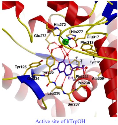
A closer look at the active site of TrpOH reveals that almost all of the listed amino acids are within the 221-360 amino acid range that is highly conserved in all three proteins.

Permission for use granted. The Stevens Laboratory Tryptophan Hydroxylase. <http://stevens.scripps.edu/tph.html>
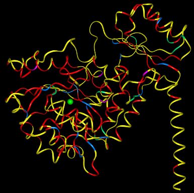
Phenylketonuria (PKU) is caused by toxic levels of phenylalanine built up in the body. Mutations in PheOH can cause this disorder. According to the Phenylalanine Hydroxylase Locus Knowledgebase, (PAHdb 2005) mutations occur in all 13 exons and flanking sequences of the gene and cause a range of phenotypes from those that are silent to those with full PKU. As of February 2, 2005, there have been 493 mutations identified. Most of the mutations occur in the catalytic domain (The Scripps Research Institute 2005 <http://stevens.scripps.edu/pah.html>.) Sequence subset 221-360 which was noted as having the highly conserved sequence, is found in the catalytic domain of the PheOH protein. Since most mutations resulting in PKU have been found in this area this again supports the theory that the catalytic domain is vital to the proper functioning of PheOH.

Permission for use granted. The Stevens Laboratory Tryptophan Hydroxylase. <http://stevens.scripps.edu/tph.html>
Point mutations are colored red, blue, magenta and green. Yellow indicates no mutation. Most of the mutations occur in the catalytic domain, while some occur in the regulatory domain.
Protein-protein Blast. 2005. Blast search (using PheOH sequence). <http://www.ncbi.nlm.nih.gov/blast/>. Accessed 2005 Mar 3.
[PAHdb] Phenylalanine Hydroxylase Locus Knowledgebase. 2005 Feb 2. PAHdb home page. <http://www.pahdb.mcgill.ca/>. Accessed 2005 Mar 7.
The Stevens Laboratory, The Scripps Research Institute. 2005 Jan 10. PKU Basic Research: Phenylalanine Hydroxylase. <http://stevens.scripps.edu/pah.html>. Accessed 2005 Mar 7.
The Stevens Laboratory, The Scripps Research Institute. 2005 Jan 10. Tryptophan Hydroxylase. <http://stevens.scripps.edu/tph.html>. Accessed 2005 Mar 7.
The Stevens Laboratory, The Scripps Research Institute. 2005 Jan 10. Tyrosine Hydroxylase. <http://stevens.scripps.edu/th.html>. Accessed 2005 Mar 7.
Contact Megan at mecastle@davidson.edu