1999 Molecular Biology Exam #3 - Using Molecular Tools
There is no time limit on this test, though I have tried to design one
that you should be able to complete within 2.5 hours, except for typing.
You are not allowed to use your notes, any books, or any electonic sources,
nor are you allowed to discuss the test with anyone until Monday May 10,
1999. EXAMS ARE DUE AT 9 AM ON MONDAY, May 10. You may use a calculator
and/or ruler. The answers to the questions must be typed on a separate sheet
of paper unless the question specifically says to write the answer in the
space provided. If you do not write your answers on the appropriate pages,
I may not find them unless you have indicated where the answers are.
Please do not write or type your name on any page other than this cover
page. Staple all your pages (INCLUDING THE TEST PAGES) together when finished
with the exam.
Name (please print here):
Write out the full pledge and sign:
How long did this exam take you to complete (excluding typing)?
10 pts.
1. Figure one shows a DNase footprint assay. R and Y are sequencing lanes
for MW standards. All protein sources are derived from mitochondiral protein.
Lane 1 used crude homogenate that was performed in a buffer containing a
lot of salt and detergent . Lane 2 used the crude homogenate after it was
dialyzed with a simple buffer. Lanes 3 9 are fractions of protein
after the homogenate was run over an affinity column matrix . Used in each
reaction are: flow through fraction of protein (lane 3), protein eluted
with 0.25 M KCl (lane 4), 0.5 M KCl (lane 5), 0.8 M KCl (lane 6), 1 M KCl
(lane 7), and 2.0 M KCl (lane 8). Lane 9 used the buffer alone and no KCl
or protein.
Interpret this figure one lane at a time and be sure to include in your
answer the following two points where appropriate:
a) the location of binding by this protein or proteins
b) how many proteins are binding here
The first two lanes are MW markers and used to
determine which bases in the promoter under investigation are bound by proteins.
Lane 1 shows that protein(s) in crude homogenate with hight salt
and detergent can bind to this DNA. It appears to be one protein binding
to the DNA in the area of bases 3256-3235 (the same location in all positive
lanes below).
Lane 2 is identical to lane one showing that this protein does not
require high salt or detergent to bind.
Lane 3 shows that the protein that binds to this DNA is retained
on the column and is not available to bind to the DNA.
Lane 4 shows the same as lane 3.
Lane 5 shows that the DNA-binding protein is eluted from the column
at 0.5 M KCl. It binds to the same location as it did in the crude extracts
in lanes 1 and 2.
Lane 6 shows the same result as lane 5 demonstrating that the protein
is still being eluted at this higher salt concentration. It hints that two
proteins might be in the crude mixture that compete for the same binding
site on the DNA, and that each protein is eluted at a different salt concentration.
Lane 7 shows that all the DNA-binding protein has been eluted from
the column with the lower salt concentrations and there is none left on
the column.
Lane 8 shows the same as lane 7.
Lane 9 is a negative control showing the banding pattern in the absence
of any added protein.
8 pts.
2. Figure 2 shows a band shift assays. They are trying to determine the
function of a Drosophila protein called extended (exd). On the left
part of the figure, they added 4 increasing amounts of engrailed protein
and no exd protein. On the right side, they added a fixed amount of exd
and the same four increasing amounts of engrailed. Interpret this figure.
From this figure we can tell that engrailed does not
bind to the probe by itself since there is no band shifted into this figure
(probe was lower in the gel). However, when a fixed amount of exd is added,
there is an increasing amount of shifted DNA probe as more and more engrailed
is added. This indicates that engrailed binds to the probe but only in the
presence of exd. Alternatively, exd binds to the probe but only in the presence
of engrailed and as more engrailed interacts with exd, more exd can bind
to the probe. Either way, these two proteins appear to require interaction
before one can bind to this DNA.
10 pts.
3. On a related topic, figure 3 shows another band shift assay. This time
they used two different probes
GTCAATTAAAGCATCAATCAATTTCG (LEFT) or
GTCAATTAAATGATCAATCAATTTCG (RIGHT).
In this experiment, they used protein combinations as indicated by the boxes.
en stands for engrailed, ubx stands for ultrabithorax, and exd you know
already.
Interpret this figure.
.exd alone cannot bind to either DNA sequence. Ubx
alone can bind to both DNA sequences. Ubx plus exd can bind as a joint complex
to both probes, but teh left probe is bound to a higher degree. en alone
can bind to both probes. en plus exd as a joint complex can bind to the
right probe but not the left probe, but en is still able to bind to the
left probe. Since the only difference between these two probes is GC (left)
and TG (right) in the middle of each oligonucleotide, we can conclude that
exd cannot bind to the right oligo by itself but with the assistance of
en, exd can bind and probably in the area of the TG difference. Also, neither
Ubx or en bind in the TG/GC areas in the middle of the probes since they
bind to both probes. Likewise, exd can bind to both probes in the presence
of Ubx and since there is a stronger band on the left gel, we have to think
this combination of proteins binds better to the GC region than the TG region.
This suggests that the exact binding domain of exd is dependent upon other
proteins modulating its shape. Alternatively, UBX and en bind to the DNA
and the sequnece differences produce different shapes in these two proteins
which influences exd's ability to bind to these two proteins.
10 pts.
4. A group of clever genetic engineers have designed a cow that produces
low lactose milk. They produced the enzyme lactase (normally secreted by
intestinal cells) in mammary glands. I would like you to design a cow to
produce low lactose milk but don't damage the meat by expressing this enzyme
in other cells. Draw a diagram or two on your answer sheet showing me what
DNA constructs you would make to generate this cow. Also walk me through
any breeding you would deem necessary.
All you would need to have is a mammary gland-specific
promoter (from a milk protein gene) upstream of a lactase gene or cDNA.
This construct could be inserted randomly in the genome or homologous recombination
could be used to produce a heterozygote. No other breeding would be necessary
other than the one to make the cows pregnant to stimulate lactation. However,
you would want to maintain some of these cows in the herd so some selective
breeding would be necessary to maintain this transgenic strain.

8 pts.
5. Figure 4 shows a large family pedigree and some molecular data related
to a genetic disease. PCR and a restriction enzyme were used on genomic
DNA, and the results electrophoresed on an agarose gel. What can you tell
me about this disease?
.This disease is due to a mutation in the DNA so that
a new restriction site is present in the mutant allele. Individuals that
have the disease have a new, lower band in the gel.
This disease is probably a dominant disease since it does not appear in
grandparents and grandchildren but the parents do not have the disease.
This disease is not sex-linked.
10 pts.
6. Figure 5 uses the CAT assay and a wide range of hormone receptors. As
you may know, most hormones are hydrophobic and their receptors are located
in the cytoplasm. When the hormone binds to its receptor, the receptor changes
shape and migrates into the nucleus and becomes a transcription factor to
activate a range of genes. Regions of promoters/enhancers that bind to hormone
receptors are called "hormone response elements". In this experiment,
they wanted to know what affect a protein called SRA would have on the ability
of hormone receptors to do their jobs as transcription factors. In the figure,
H = hormone. It is interesting to note the use of RU486, the "morning
after pill".
a) Tell me what affect SRA has as revealed in this experiment.
.SRA enhances the activity of these hormones: P, G,
A, E, a little bit for thyroid hormone, but not at all for the last 3 hormones.
We also learn that SRA cannot activate the PR by itself, nor in the presence
of RU486.
b) What is the function of RU486 in this experiment?
.RU486 acts as a control since it binds to and inactivates
the receptor for hormone P. It serves as a
negative control.
34 pts.
7. Three figures for this question can be found on the web at this URL:
<Restricted
Access - Campus Only>. To view it, type in the URL, then print the
figures but make sure you do not leave prints all over campus in case someone
in class finds it who has not started taking the test. They are from a paper
that was examining a very important organ that most people do not even know
exists the vomeronasal organ (VO) which is what detects pheromones
in humans (I'm not making this up). VR i2 is a particular receptor in VO
neurons used to detect pheromones and the researchers are trying to make
transgenic mice.
a) Read carefully the figure legend that appears on page 4 of this test.
The black boxes represent noncoding (i.e. 5' and 3' UT). The IRES section
permits the two genes (VR i2 and the reporter gene) to be transcribed as
a single mRNA but each protein to be translated separately. Finally, a fusion
protein of tau and reporter will result from some of these constructs. The
tau portion of the reporter genes causes the fusion proteins to bind to
all microtubules, which labels the full length of the axons (www figure
3).
Tell me how you would be able to tell which cells contained the DNA shown
in lines a, c, d, e, f, and g (www figure 1). You cannot use PCR.
.I would use RFLP analysis to determine which construct
was present in cells. For each construct, I would isolate genomic DNA and:
Line a: cut with HindIII and probe with a probe containing the VR2
coding region. This would produce a band of 8.5 kb.
Line c: cut with HindIII and probe with a probe containing the VR2
coding region. This would produce a band of 17.3 kb.
Line d: cut with HindIII and probe with a probe containing the VR2
coding region. This would produce a band of 14.8 kb.
Line e: cut with HindIII and probe with a probe containing the VR2
coding region. This would produce a band of 11.6 kb.
Line f: cut with HindIII and probe with a probe containing the GFP
coding region. This would produce a band of 14.6 kb.
Line g: cut with HindIII and probe with a probe containing the M71
coding region. This would produce a band of 14.6 kb.
b) What is the difference between the DNA in lines c and d? How was this
difference generated and in what cells would it be done?
The difference is that the neomycin resistance gene
has been deleted by a Cre-loxP approach. This would happen only in cells
that produced Cre, which is controlled by its promoter. It might have happened
in the stem cells since neo selection is not needed after that point.
c) The purpose of this experiment was to create different reporter gene
constructs so that mice would be heterozygous for constructs VG and VL (www
figure 1). Tell me how you would make such a mouse given these constructs.
You do not need to list every manipulation, but do tell me which
cells and mice and major procedures you would use to generate these mice.
Do not duplicate your answer from part b above. Mice containing constructs
f and g were used as controls, but we will not consider those here.
You must make a homologous recombination vector that
allows you to do the Cre-loxP cell-specific recombination (line b and GFP
equivalent - exchange lacZ for GFP) as well as one for GFP.
You must each construct separately into stem cells via homologous recombination
(line c and GFP equivalent) and select for neo resistance.
You must delete the neo marker since it is no longer needed (lines d and
e). Cre must be produced in these cells - any strong promoter in the stem
cells would do and the Cre could be transiently expressed.
These stem cells would need to be put into a blastocyst and mosaic mice
produced - one for lacZ and one for GFP.
These mice would be mated to produce heterozygous (mutated and wt) mice
that are 100% transgenic - one strain GFP and one strain lacZ.
These mice would be mated to produce mice that are heterozygous for the
GFP construct and the lacZ construct.
d) In this experiment, they wanted only neurons from the vomeronasal
organ to express the tau-reproter fusion proteins. How was this accomplished?
They relied upon the fact that the IRES portion permitted
on mRNA to be translated as two separate proteins. This allowed them to
use the normal VR2 promoter and enhancer since homologous recombination
was used.
e) They generated these VG/VL mice, and found that vomeronasal neurons
expressed either GFP or ß-galactosidase, but never both (www figure
2). Interpret these results.
This tells us that in VO neurons, there is some form
of allelic exclusion going on such that one allele is expressed but the
other is silent. This means in normal mice, any given VO neuron would express
only one type of receptor, and not a mixture of two receptors. This would
allow each neuron to respond to only one of two possible ligands.
10 pts.
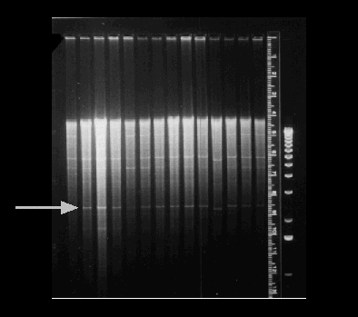
8. The last question... Determine the molecular weight of the band indicated
by the arrow. The standards on the right side of the gel are in kilobases.
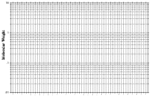
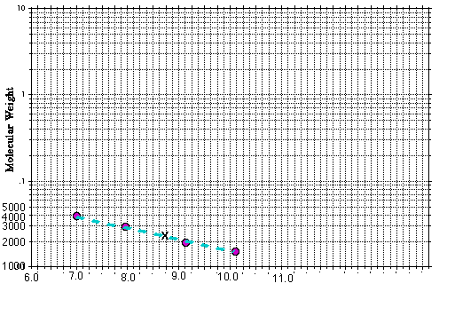
You MUST use the graph provided here and put your answer in the blank below.
The answer is about 2300 bp.


Answer graph

Return to exam without
answers
Return to Molecular
Main Page
Return
To Biology Main Page

© Copyright 2000 Department of Biology,
Davidson College, Davidson, NC 28036
Send comments, questions, and suggestions to: macampbell@davidson.edu


