This web page was produced as an assignment for an undergraduate course at Davidson College.
Microbiota and Growth in
Mice during Chronic Undernutrition - Summary
Summary:
In this paper, Schwarzer et
al. provide evidence that the intestinal microbiota sustains
weight gain and longitudinal growth in mice as well as interacts with
the somatotropic axis to sustain systemic growth. They subjected wild
type (WT) and germ free (GF) juvenile mice to a standard and
nutritionally depleted diet. They found that when fed a standard diet,
GF mice weighed less and were shorter. When weaned onto a nutritionally
depleted diet, both WT and GF mice lost weight, but GF mice lost more
weight. After readaptation to solid food, WT mice resumed growth and
recovered weight and longitudinal growth while GF mice did not. They
studied the effects of the microbiota on the somatotropic axis by
measuring levels of IGF-1 (insulin-like growth factor-1) and GH (growth
hormone). Several measurements indicated that GF mice had lower levels
of GH and IGF-1 compared to WT mice. To further test this, they injected
both WT and GF mice with recombinant IGF-1 and found that as a result,
GF growth levels increased to levels seen in WT mice. They also treated
mice with picropodophyllin (PPP), a specific noncompetitive inhibitor of
IGF-1R that delayed the growth of WT mice. This provided more evidence
that IGF-1 activity is important to the growth of WT mice.
Finally, Schwarzer et
al. identified a specific strain of the bacteria Lactobacillus
plantarum that counters the GH resistance in mice caused by
chronic undernutrition. Two strains of this bacteria were monocolonized
in mice that were subjected to chronic undernutrition: one (LpWJL)
resulted in more weight gain and bone growth than the other (LpNIZO2877).
Collectively, their data showed that juvenile growth in mice is affected
by the gut microbiota, specifically a strain of Lactobacillus
plantarum, that contributes to the maintenance of growth during
chronic undernutrition.
Opinion:
This paper provides evidence from many
experiments in support of the important role of the intestinal
microbiota in juvenile growth in mice. Each experiment fit well into the
story they constructed and helped to confirm the basis of their claims.
For example, when providing evidence for the microbiota’s effects on the
somatotropic axis, they supported their claims with data from a variety
of measurements: GH, IGF-1, and IGFBP-3 levels; expression of GH-receptor,
Igf1, Igfbp3,
and Socs3;
phosphor-S473-Akt signal. It would have been interesting to see the
actual breakdown of WT gut microbiota to see how much of the WT
microbiota consists of the mentioned lactobacilli strains. Additionally,
it would have been helpful if figure 3 and figure 4 had been switched in
the order they appeared since figure 4 was referenced before figure 3.
Summary
of Figures:
Figure
1
summarizes the effects of the microbiota on mice fed a standard breeding
diet. Weight (A) and body length (C) of juvenile mice over time is shown
to be significantly different, especially after weaning. The increases
in weight gain and body length per day (B and D) are also significantly
different as a result of the microbiota. Bone growth parameters such as
femur length (E) and cortical thickness (F) were significantly lower in
GF mice than WT mice.

Figure 2 summarizes results from various methods of support for the role of the microbiota on the somatotropic axis and its effects on growth of juvenile mice. Levels of GH, IGF-1, and IGFBP-3 (A, B, and C, respectively) in the sera of mice show that GH levels gradually decreased during postnatal growth, IGF-1 was consistently reduced in GF mice while shooting up in WT mice at 28 days, and IGFBP-3 was also consistently and significantly reduced in GF mice at the time points tested. Igf1 and Igfbp3 (D and E, respectively) expression was measured at 28 days, the same time that IGF-1 levels peaked in WT mice, and was found to have reduced expression in GF mice. Phosphorylation of Akt, another measure of IGF-1, was found to be reduced in the liver of GF mice (F).

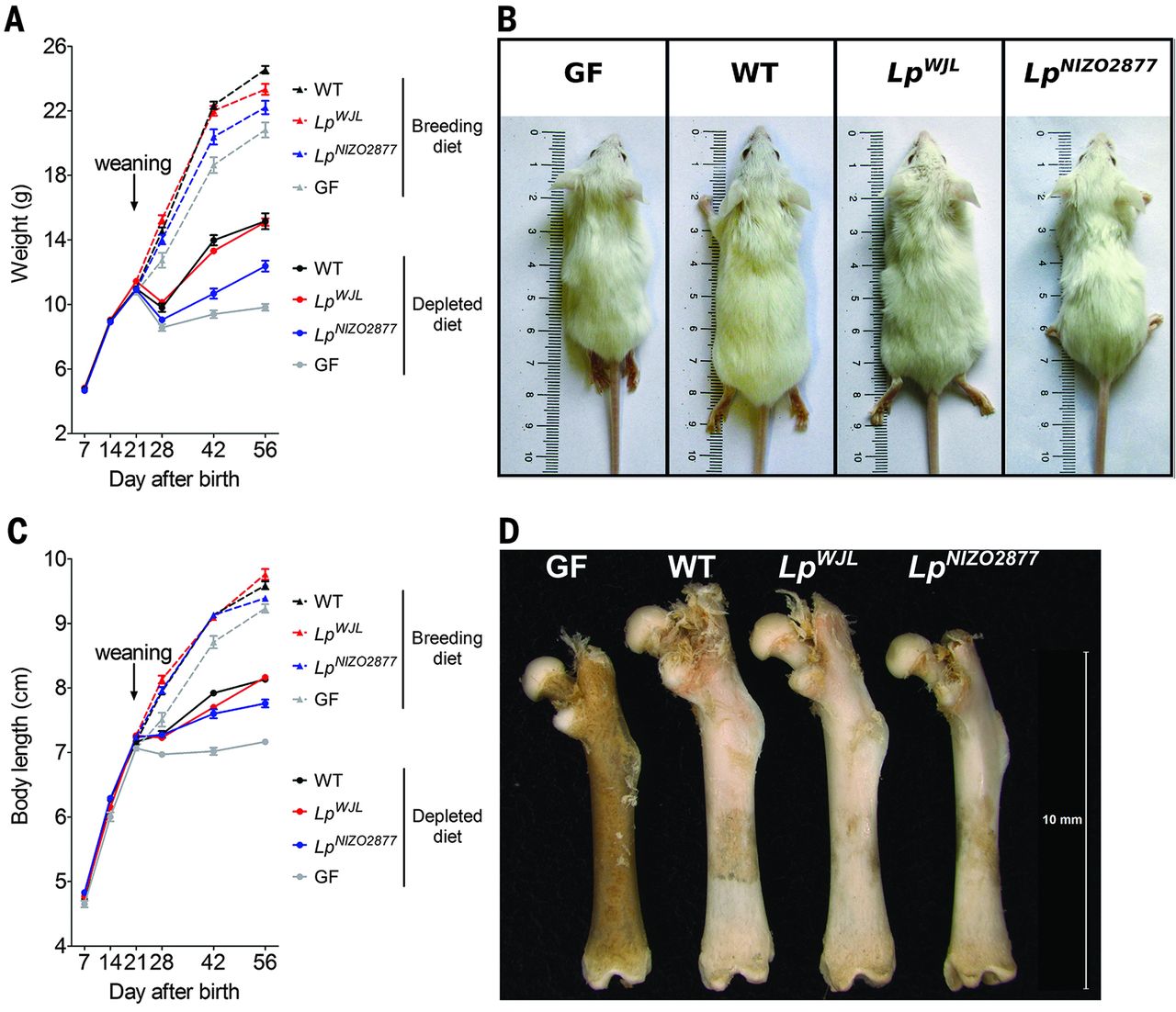
Figure 3 compares weight and body length of WT, GF, and GF monocolonized with either LpWJL or LpNIZO2877 after being fed either a standard or nutritionally depleted diet (A and C). After weaning onto their respective diets, mice on the nutritionally depleted diet weighed significantly less and were significantly shorter than mice on the standard diet. Within each diet condition, WT and LpWJL monocolonized mice had similar weight and body length while GF and LpNIZO2877 monocolonized mice weighed significantly less. Differences in body length and femur length for each of the conditions of mice fed a depleted diet are shown (B and D, respectively).

Figure
4 shows
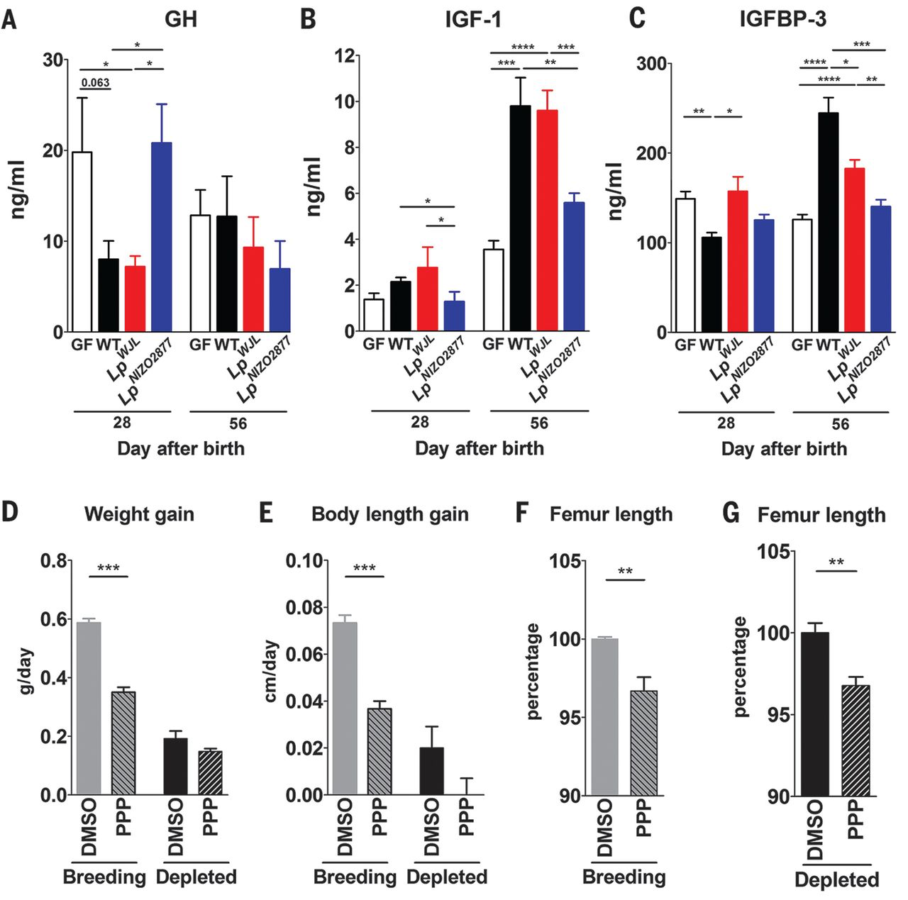
the levels of GH (A), IGF-1 (B), and IGFBP-3 (C), as a result of chronic
undernutrition on WT, GF, GF monocolonized with LpWJL, and GF monocolonized with LpNIZO2877 mice 28 and 56 days after birth. GH levels in
the sera were significantly increased in GF and LpNIZO2877-monocolonized mice after 28 days while IGF-1
and IGFBP-3 were significantly increased in WT and LpWJL-monocolonized mice after 56 days. Panels D-G show
that when compared to DMSO (control), WT mice treated with PPP, a
noncompetitive inhibitor of IGF-1, had significantly less weight gain
and body length gain in mice fed the standard diet, and significantly
reduced femur length in mice fed both diets.

Reference:
Schwarzer, M., Makki K., et al.
2016. Lactobacillus plantarum strain maintains growth of infant mice
during chronic undernutrition. Science
351(6275):854-857. Web.
Email Questions or Comments: saayyar@davidson.edu.
© Copyright 2016 Department of Biology, Davidson College, Davidson, NC 28035