This page will help you learn how to get the most from chime, a free plugin for viewing 3D structures.
Before you begin, you might want to see the differences between Mac and PC mouse controls. The directions below are written for Macs, but the differences are minor.
If your chime plugin is working, click here and see the 3D structure of a transcription factor (called Cro) binding to DNA.
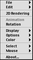
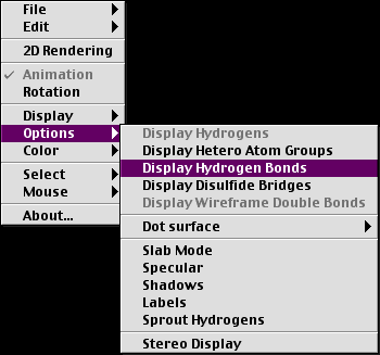
At first, the structure won't look like much. To help you see what's what, click and hold on the structure until the window appears:
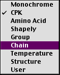
Mouse down on this window while still clicking and go to the color option and choose "Chain". The check indicates that the CPK color scheme is currently in use.
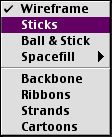
Next, Click and choose the Display menu, and select "Sticks":
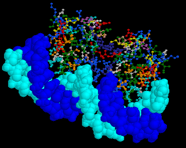
Click and drag around the structure until your view looks something like this:
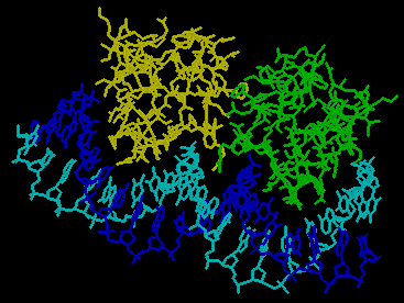
You can see that this structure is made of 4 chains (one for each color). The DNA two strands are two shades of blue while the protein is revealed to be a dimer (one yellow and one green). Now hold down the shift key and click and drag the mouse so that you can zoom in (mouse down) and zoom out (mouse up). Try to create a view like this:
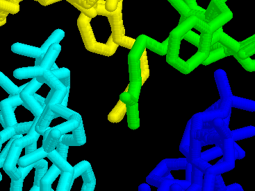
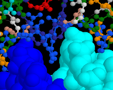
You can see two amino acids protruding into the minor groove of the DNA. You might need to shift the structure around a bit to see this exact view. To do this, hold down the option key and click and drag the mouse. See if you can find one end of this DNA, the side with the yellow protein binding:
Now, zoom back out and recenter the structure. Now let's look at some other views. Choose structure from the color menu and ribbon from the display menu. You should see something like this:

You can see that the DNA does not have any color since structure indicates secondary structure for proteins only. However the ribbon display shows red alpha helices on the protein (beta sheets would appear yellow) and white portions have no secondary structure. It is easier to see the major and minor grooves of DNA with this view.
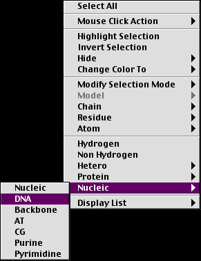
Let's try another some new options. Choose the select menu and then choose Nucleic and DNA from the subsequent menu:
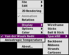
From the display menu, choose Spacefill and Van der Waals Radii from the subsequent menus.
From the color window, choose chain again.
Now in the select menu, choose protein. Then choose protein:

And then go to the display menu and select "Ball & Stick". In the color menu, choose "Amino Acid". You should have a view similar to this:
Now go to the select menu, and choose "Select All". Go to the "Options" menu and choose "Display Hydrogen Bonds":
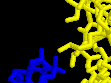
When you do, you will see the many hydrogen bonds wihtin the protein. Zoom in on the minor groove to see if any hydrogen bonds (dotted lines) have formed between the DNA and the protein. Try to get a view similar to this:
As you can see, there are no hydrogen bonds here. Use the mouse to click on the very end of the light blue amino acid that is protruding into the minor grove. When you do, Chime will identify what you have clicked on. The information appears at the bottom left corner of the window:
![]()
From this information, you can see that you have clicked on arginine (which carries a postive charge), which is amino acid #43 on chain R (the letter R is not informative and seems to be assigned randomly). You have also clicked on a nitrogen which is atom number 1745 in this chain.
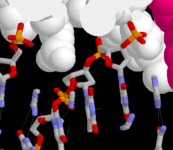
Let's try one more neat trick before you explore on your own. From the options menu, choose "Slab Mode". Slabbing allows you to peel away layers of a structure so you can peak inside. When you did this option, you should have seen some of the structure disappear. It is just hiding from view but if you rotate the structure, it will come back into view but other parts will disappear. Try moving the structure around some. To change the depth of slabbing, hold down the control button while clicking and dragging the mouse up and down. Play with these controls and slice into the protein and DNA.
I have changed the settings for color and display and then slabbed in to see what parts of the DNA are binding to the protein (structure color and space fill display). At this location, you can see the phosphates portruding out towards the protein. You can also see the hydrogen bonds between base pairs of the DNA.
Given this view and the amino acids sticking into the minor groove how do you think the protein and DNA adehere to one another?
Now you have seen some of what chime can do. Explore all the options and you will discover chime can provide you with even more information.
![]()
![]()
© Copyright 2001 Department of Biology, Davidson College, Davidson, NC 28036
Send comments, questions, and suggestions to: macampbell@davidson.edu