Simple Squamous Epithelium
Simple squamous epithelial cells are thin and flat (the thinnest of all epithelial cell-types), which allows them to have a large surface area that is exposed to the lumen on one side (the apical surface), and to the basement membrane (i.e. basal lamina) on the other (the basolateral surface; Figure 1). The cells, scale-like in appearance, tend to have larger, elliptically-shaped nuclei. As a simple type of epithelium, simple squamous epithelium is one cell-layer thick, and thus every cell of the tissue comes in direct contact with the basement membrane. As with other types of epithelia, simple squamous epithelial cells are bound together by tight junctions, forming a selective barrier, which is crucial to its function. |
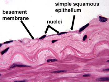
Figure 1: Hand-drawn cross section of simple squamous epithelial tissue (not to scale). |

Simple squamous epithelial cells function as mediators of filtration and diffusion. Due to their simple and thin construct, they allow for easy transmembrane movement (i.e. across the membrane, and through the cell) of small molecules. Some molecules, such as oxygen and carbon dioxide diffuse freely across the simple squamous epithelia according to concentration gradients. Other molecules, such as ions, utilize transmembrane protein channels to diffuse across the cells; therefore, the types of proteins that are present in a given simple squamous epithelial tissue paritally determines the function of that tissue. Therefore, the simple squamous epithelia helps to determine what is able to move from the lumen and into the bloodsteam (the capillary bed is under the basement membrane), and vice versa.
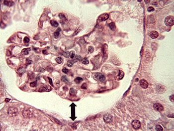
| As could be expected, simple squamous epithelium is found in locations where rapid diffusion or filtration take place. In the kidney, simple squamous epithelium lines the Bowman’s capsule and the glomerulus (Figure 2). In this case, the ability of simple squamous epithelium to provide rapid filtration and diffusion is instrumental to the kidney's function, as the kidney filters about 180 liters of blood per day. |
Figure 2: Simple squamous eplithelia in the kidney. The arrow pointed upwards is directed toward the glomerulus, the downward arrow is directed toward the Bowman's capsule; both are lined with simple squamous epithelial tissue. |
| Simple squamous epithelium also makes up the alveoli in the lungs (Figure 3), where rapid gas exchange occurs, as oxygen enters the bloodstream through alveolar blood vessels and carbon dioxide exits as waste. |
Figure 3: The alveoli of the lungs, which are made up of simple squamous epithelial tissue (labeled alveolar epithelium in the image). |
In addition, simple squamous epithelium lines the cavities, which arise from the coelom of the mesoderm (i.e. the peritoneal, pleural and pericardial cavities); simple squamous epithelium is referred to as “mesothelium” in these cases. “Endothelium,” which lines the interior surfaces of the entire circulatory system is another example of a specific form of simple squamous epithelium (Figure 4). |
Figure 4: Endothelial tissue of a blood vessel. |
References
Ackerley, S.K. (2005, December 9). Epithelial Tissue. Retrieved from
http://www.uoguelph.ca/zoology/devobio/210labs/epithelial1.html
Alberts B., Johnson A., Lewis J., Raff M., Roberts K., & Walter P. (2002). The Molecular Biology of The Cell (4th ed.). New York, NY: Garland Science.
Epithelium.(2010). Retrieved from http://ect.downstate.edu/courseware/histomanual/epithelia.html.
Finazzo, S. (2009). Epithelial Tissue. Retrieved from http://facstaff.gpc.edu/~sfinazzo/epithelium%20edited/epitheliumIndex.html.
King, D. (2010, April 13). Epithelium Study Guide. Retrieved from http://www.siumed.edu/~dking2/intro/epith.htm#simpsquam.
Tamarkin, D.A. (2006). Glomerular Filtration. Retrieved from http://faculty.stcc.edu/AandP/AP/AP2pages/Units24to26/urinary/filtrati.htm.