Cell Polarity
In cells, polarity refers to the asymmetric organization of different aspects of the cell including the cell surface, intracellular organelles and the cytoskeleton. These characteristics are then maintained through cell division. Differentiation in the ways these parts are asymmetrically organized helps to determine cell function and cell type. In epithelial cells, the individual cells are split into two regions, the apical and basolateral regions, which are chemically and structurally different from each other (Figure 1). The apical region is defined as the area lying above the tight junctions (to learn more about tight junctions, click here) and contains the apical membrane which faces the lumen or the outer surface. The basolateral region is the side that is below the tight junctions and contains the basolateral membrane which is in contact with the basal lamina. In addition, epithelial cells have lateral membranes, which are often characterized by tight junctions, gap junctions, and other cell-cell interactions which help to form epithelial tissue. |
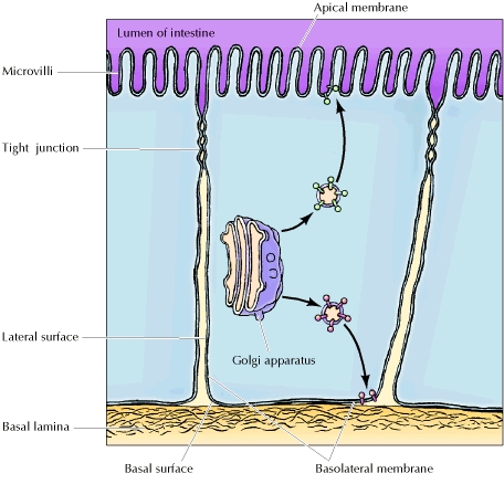 Image from: http://www.ncbi.nlm.nih.gov/bookshelf/br.fcgi?book=cooper&part=A1498 Figure 1.Side view of an epithelial cell, displaying apico-basal polarity |
Epithelial Morphogenesis
| Polarization is also instrumental in the morphogenesis of epithelial cells (figure 2). At the 8-cell stage, e-cadherin is activated through phosphorylation. At the 16-cell stage, e-cadherin recruits the actin cytoskeleton and associated proteins (i.e. spectrin, ankyrin, fodrin), which allow for binding and movement of membrane proteins (e.g. Na+/K+ ATPase). In addition, e-cadherin recruits the Sec6/Sec8 protein complex, which serves as a docking receptor for vesicles sent from the Golgi apparatus to the basolateral membrane. This allows for the polarization of membrane proteins and pumps, which contributes to the formation of concentration gradients in the forming embryo. These concentration gradients are instrumental in further cell differentiation. |
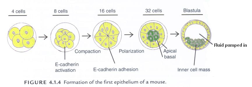 Image from: Davies, J. A. (2005). Mechanisms of morphogenesis. Burlington, MA: Elsevier Inc. Permission pending. Figure 2. Side view of an epithelial cell, displaying apico-basal polarity |
Polarity is maintained through a set of three orchestrated mechanisms. Different combinations of these mechanisms lead to different polarization in various types of epithelial cells.
- Selective packaging of proteins – epithelial cells use distinct carrier vesicles and sorting by the trans-Golgi network (TGN) in order to package proteins and send them to either the apical or basolateral surface.
- Selection and depletion after random delivery – proteins are sent to both the apical and basolateral surfaces in equal amounts. However, some proteins are more stable on one surface than the other; meanwhile, others are unstable and broken down. Therefore, there is a gradual construction of differential concentrations of proteins on each surface.
- Transcytosis – To learn more about this mechanism, see the Transcytosis page.
Differentiation within epithelial cells
|
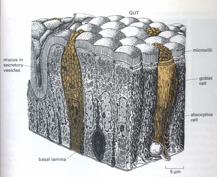
Polarity varies according to cell type and leads to different functional usage. One example of this differentiation can be seen in goblet and absorptive cells in the small intestine (Figure 3). Although both types of cells are classified as simple columnar cells, their differing polarity results in their different functional capabilities. Goblet cells main role is to produce mucus to protect and lubricate the gut. The asymmetrical organization of the cells’ Golgi apparatus, secretory vessels and cytoskeleton enable them to discharge the mucus from their apical surface, which is the surface lining the gut. Absorptive cells, on the other hand, must import food molecules through their apical surface, before exporting the cells through their basal surface into the underlying tissue. In each case, the apical and basolateral parts of the membrane are doing two different sets of functions, which require them to have different sets of membrane proteins. To learn how they get these different proteins, see our Maintenance of polarity & regulation of membrane trafficking section. |
Which types of cells are polar?
Epithelial Cells |
As explained in the section “What is Polarity?” epithelial cells display apico-basal polarity, as well as specialized lateral membranes which allow for specialized diffusion/absorption of ions, secretion, and adhesion between cells (Figure 4). |
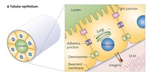 Image from: Bryant, D. M., & Mostov, K. E. (2008). From cells to organs: Building polarized tissue. Nature Reviews Molecular Cell Biology, 9(11), 887-901. Permission pending. Figure 4. Polarization in epithelial cells. |
Immune System Cells |
Migratory cells of the immune system, such as neutrophils, use polarization for short-distance communication. They display strong front-back asymmetry and demonstrate polarized migration towards attractive cues (i.e. chemotaxis), such as Interleukin-8. |
|
Neurons |
Polarity in neurons is necessary for long range communication. Polarization is utilized in passing of electronic signals through neurons, which help to connect the Central and Peripheral Nervous Systems (CNS and PNS). In addition, polarization is used in the targeting and uptake of neurotransmitters (Figure 5). |
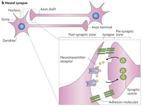 Image from: Bryant, D. M., & Mostov, K. E. (2008). From cells to organs: Building polarized tissue. Nature Reviews Molecular Cell Biology, 9(11), 887-901. Permission pending. Figure 5. Polarization in neuron cells. |
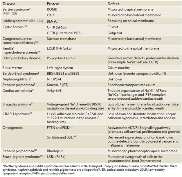
What happens when polarization goes wrong?
References
Alberts, B., Bray, D., Hopkin, K., Johnson, A., Lewis, J., Raff, M., et al. (2004). Essential cell biology (2nd ed.). New York, NY: Garland Science.
Bornens, M. (2008). Organelle positioning and cell polarity. Nature Reviews Molecular Cell Biology, 9(11), 874-886.
Bryant, D. M., & Mostov, K. E. (2008). From cells to organs: Building polarized tissue. Nature Reviews Molecular Cell Biology, 9(11), 887-901.
Childs, G. V. (2002). Medical cell biology epithelia study guide. Retrieved 9/26, 2010, from http://www.cytochemistry.net/microanatomy/epithelia/epith_lec.htm
Cooper, G. M. (2000). The Cell, A Molecular Approach (2nd ed.) Sunderland, MA: Sinauer Associates. Reprinted online at: <http://www.ncbi.nlm.nih.gov/bookshelf/br.fcgi?book=cooper>
Davies, J. A. (2005). Mechanisms of morphogenesis. Burlington, MA: Elsevier Inc.
Mellman, I., & Nelson, W. J. (2008). Coordinated protein sorting, targeting and distribution in polarized cells. Nature Reviews Molecular Cell Biology, 9(11), 833-845.
Pollard, T. D., Earnshaw, W. C., & Lippincott-Schwartz, J. (2008). Cell biology (2nd ed.). Philadelphia, PA: Elsevier Inc.