A Proteomic Look at STM1 and YLR152C

This web page was produced as an assignment for an undergraduate course at Davidson College.

Review and Purpose

After sequence analysis and microarray expression profile analysis, I supported the prediction that the annotated gene STM1 works in translation as well as in telomere maintenance and anti-apoptosis. My findings on the non annotated gene YLR152C led to the predictions that YLR152C functions as a membrane protein that either provides transport for molecules in metabolic pathways or works directly in a metabolic pathway while attached to some membrane, possibly the mitochondria. By examining each proteins' expression and interactions, I will reevaluate my earlier predictions and propose future experiments that will test further predictions.

Note: Structural analysis of the proteins could not be provided as currently neither protein has been crystallized.

STM1

2D Gel  

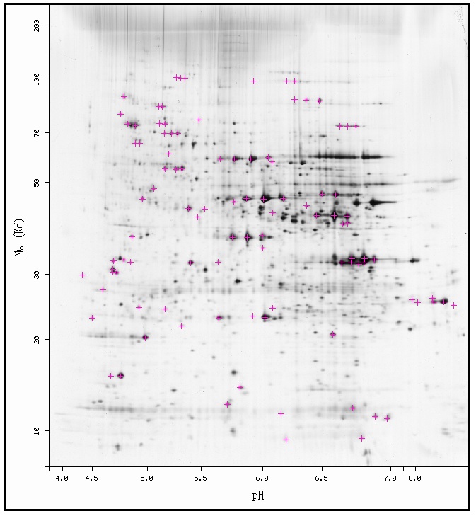
By using PROWL's PROTEININFO section, I discovered that STM1 has a molecular mass of 29977.260 Da and an isoelectric point of 9.7 (PROWL PROTEININFO 2006, http://prowl.rockefeller.edu/prowl-cgi/ReadSequence.exe?name=db|1|nr-Saccharomyces-cerevisiae|gi|6323179|ref|NP_013251.1|). I then compared that information to the 2D-PAGE gel of yeast proteins and found that STM1 has a isoelectric point larger (more basic) than what is provided in the gel below ( ExPASy Swiss-2DPAGE Viewer, http://ca.expasy.org/swiss-2dpage/viewer?map=YEAST&ac=all)

From this information, I found that in order to isolate STM1 using 2D gel electrophoresis a gradient that has better sensitivity for proteins with high isoelectric points is necessary; thus, experiments using this technique must take the isoelectric point and senstivity of the gel into consideration.


Protein interactions  

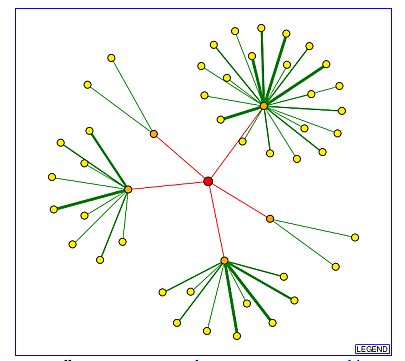
The figure below is a node graph for the interactions of STM1. STM1 is represented by the red node in the center. The orange nodes interact directly with STM1 while the yellow nodes interact directly with its attached orange node. The red lines indicate that while five interactions have been noted for STM1, the experiments that led to their discovery were high-through put and other means have yet to be employed to confirm the results.

(DIP 2006, http://dip.doe-mbi.ucla.edu/dip/Search.cgi)

The list below contains STM1 and the proteins for the five orange nodes seen above with their functions (DIP 2006).

Protein Function Location
STM1 Telomeric DNA binding cytosolic ribosome, cytoplasm, nuclear telomere cap complex
Sla1p protein binding, cytoskeletal protein binding actin cortical patch
YJR072C protein binding cytoplasm
Mec3p DNA binding nucleus
Sec31p structural molecule activity COPII vesicle coat
Hek2p mRNA binding cytoplasm, nuclear chromosome, telomeric region

Using BIOGRID, I searched for STM1 to see if the above interactions had any more data. Through BIOGRID, I found interactions listed for Sla1p, Mec3p, Sec31p, and Hek2p as well as the tests used to confirm the interactions. Below are the entries for the above mentioned proteins. Interestingly, BIOGRID lists a total of 106 proteins with associations to STM1. While no node graph or degree of association is listed, later use of BIOGRID in searching for YLR152C led me to the conclusion that the interactions are directly with STM1 as the listing for YLR152C did not include any proteins that were in the second shell of the DIP node graph (To see the full BIOGRID list please click here).The affinity capture part of affinity capture - mass spectrometry seems to be very similar to the first part of immunoprecipitation (SGD 2006, http://www.yeastgenome.org/help/glossary.html).

(BIOGRID 2006, http://www.thebiogrid.org/SearchResults/summary/31419)

Most of the interactions above support the known and predicted information on STM1. Hek2p and Mec3p are proteins that bind to nucleic acid and are in the same region as STM1. Their locations and functions support STM1 working in the telomeric and anti-apoptosis processes that are recognized in its Gene Ontology. Sla1p and YJR072C along with Hek2p support the predicted translational function of STM1. These three proteins bind protein or mRNA, which are both part of the translational pathway either as a starting material or end product, would be likely to interact with STM1 if it were helping with translation especially as Hek3p and YJR072C share cellular locations with STM1. While DIP lists these interactions as being found with high through-put methods, the affinity capture experiments sited at the BIOGRID website seem more firm and less like high through-put methods that need strong confirmation. This is interesting as Sec31 works as part of the vesicle. The interaction could come about as STM1 is present in the cytoplasm and seems to show both nucleic acid and protein binding capabilities. The Hek2p, Mec3p, and YJR072C as well as most of the other 106 interactions listed on BIOGRID support STM1's part in telomere maintenance, anti-apoptosis, and translation.

mTn Disruption  

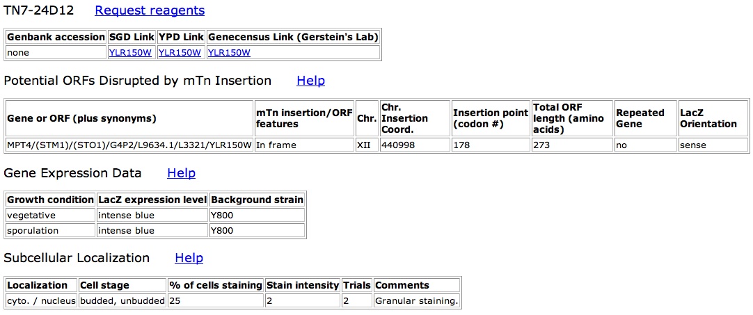
STM1 had 5 successful in-frame mTn insertion clones (seen below).

(TRIPLES 2006, http://ygac.med.yale.edu/triples)

Four of these five clones showed intense blue/strong lacZ expression during both sporulation and vegetative growth conditions while TN7-54A3 showed only faint blue/lacZ expression. Also TN7-24D12 actually showed some localized expression in the nucleus as well as the whole cell. (The pages for TN7-54A3 and TN7-24D12 are shown below. TN7-24D12's Gene expression data is being used to represent what was seen for the gene expression data of all clones that expressed intense blue).


(TRIPLES 2006, http://ygac.med.yale.edu/triples)

The knockout data suggests a STM1 has a relatively strong promoter as in most cases lacZ showed intense expression when in frame. This lacZ expression hints at a strong expression of STM1 as well. The localization of staining in clone TN7-24D12 in both the nucleus and the whole cell supports the formal gene ontology for cellular component, which places STM1 in the nucleus, cytosolic ribosome, and cytoplasm.

Conclusions and Further Experiments  

Most of the proteomic data provides further support for the earlier predictions from structural and microarray analysis. As the most interactions occurred between other DNA binding or translational pathway proteins, I predict that the theory presented by Van Dyke et al. 2004, STM1 has responsibilities in the translational pathway as well as in telomere maintenance and anti-apoptosis, holds true. The staining in the nucleus and cytoplasm by TN7-24D12 supports the claims of the current gene ontology as well as lending plausibility to my previous prediction because all the areas where the processes occur seem to express STM1.

The first experiment that I would suggest is to crystallize STM1 and determine its structure. As it is not a membrane protein and seems to have decent expression throughout the cell, STM1 seems to have the potential to be crystallized. Structural analysis has brought to light new information on binding sites in other proteins; that type of information could prove very useful while trying to determine how STM1 works in translation as well as telomere maintenance and anti-apoptosis.

The second experiment that I would suggest is using isotope-coded affinity tags to compare protein expression under the conditions that showed major repression or induction of RNA for STM1with microarrays. As seen in the gene expression website for STM1, multiple conditions produced great repression or reduction. A threshold value of induction and repression could be set. All conditions that produced values greater than the threshold value would undergo the ICAT method. In the end we would have quantified the repression or induction of STM1 protein for those times. We could then compare the protein expression with the RNA expression for the protein. If the two expressions are not the same (RNA shows an induction while the protein remains stable or is repressed and vice versa), we could run further experiments to see if STM1 is not being activated by phosphorylation or other methods or if it is being activated more efficiently than normal.

While doing the ICAT experiments based on microarrays, we can also check for the change in expression throughout the cell cycle. What a protein does at different times can be very telling about its actual function and purpose; therefore, gathering this information will expand our knowledge of STM1.


YLR152C

2D Gel  

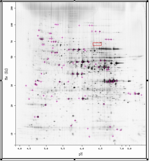
By using PROWL's PROTEININFO section, I discovered that YLR152C has a predicted molecular mass of 63990.777 Da and an isoelectric point of 6.4 (PROWL PROTEININFO 2006, http://prowl.rockefeller.edu/prowl-cgi/ReadSequence.exe?name=db|1|nr-Saccharomyces-cerevisiae|gi|6323181|ref|NP_013253.1|). I then compared that information to the 2D-PAGE gel of yeast proteins. In the figure below, a red box higlights the area where YLR152C is thought to be located ( ExPASy Swiss-2DPAGE Viewer, http://ca.expasy.org/swiss-2dpage/viewer?map=YEAST&ac=all).

While it has not been identified on the gel, several light staining dots could represent this protein. If researchers employed mass spectrometry on this section of gel, we could determine whether YL152C actually has the size and isoelectric point that we predict.


Protein interactions 

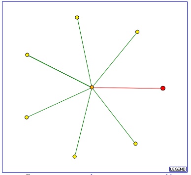
The figure below is a node graph for the interactions of YLR152C. YLR152C is represented by the red node at the right of the graph. The orange node in the center interacts directly with YLR152C while the yellow nodes interact directly with the center node. The red line indicates that the experiments that noted this interaction was high-through put and other means have yet to be employed to confirm the results.


(DIP 2006, http://dip.doe-mbi.ucla.edu/dip/Search.cgi)

Interacting Protein Function Location
Chk1p protein kinase activity nucleus

I once again used BIOGRID to check for more information on the above interaction. Chk1p was once again the only protein listed as associated with YLR152C. the process used to determine this interaction was affinity capture -MS.

(BIOGRID 2006, http://www.thebiogrid.org/SearchResults/summary/31421)


As investigators detected only a single interaction, the information is hard to use on its own to support or refute earlier predictions. The interaction with a protein kinase does not rule it out as a membrane protein as some membrane proteins are phosphorylated to activate a channel or other types of membrane proteins. As researchers performed affinity capture - MS and not yeast-two-hybrid, a stonger possibility exists that YLR152C is present in the nuclear membrane.

mTn Disruption  

No information existed for YLR152C in the TRIPLES Database.

Conclusions and Future Experiments  

The proteomic information available for YLR152C is sparse. The limited information points towards the protein being found in the nucleus as it was found to interact with a protein kinase, which is located in the nucleus. As the experiment that found this interaction was an affinity capture-MS and not a yeast-two-hybrid, I have more confidence that the protein could exist in the nuclear membrane. As no proteomic data currently refutes the prediction that YLR152C is a membrane protein and Kyte-Doolittle Hydropathy plots predict at least four transmembrane domains (see my favorite yeast gene), more information is needed before we can reject the hypothesis that YLR152C is a membrane protein. The current proteomic data does not seem to support YLR152C working directly in one of the metabolic pathways like glycogen synthesis.

The first experiment that I would suggest is the identification of YLR152C by mass spectrometry. While the 2D-gel had faint spots where YLR152 is predicted to be, YLR152C has yet to be identified and may not actually exist there. If a regular 2D-gel does not contain YLR152C in its predicted area, a second gel should be run using the detergent Triton X-114. Triton X-114 keeps membrane bound proteins suspended in it and allows a 2D-gel to resolve the proteins; thus, YLR152C would probably be resolved using this method if the prediction that it is a membrane protein holds true.

As the only known interaction is with the protein kinase in the nucleus, attempts should be made to find the subcellular localization. I suggest creating mTn disrupted clones. The cells can then be stained to see if there is any localization as has been done with other proteins found in the TRIPLES database. The localization would at least tell if YLR152C does indeed stay in the nucleus and may also be able to discern whether YLR152C exists in the nuclear membrane or in the nucleus at large.

As with STM1, we can gain great knowledge if we perform experiments that check actual protein expression against mRNA expression for the protein. By using the ICAT method with cells grown under conditions that show an induction or repression of mRNA greater than or equal to a threshold value or even under conditions that show any change in expression, we could find what happens to the protein expression for the same conditions. If the two expressions are not the same (RNA shows an induction while the protein remains stable or is repressed and vice versa), we could run further experiments to see if YLR152C is not being activated by phosphorylation or other methods or if it is being activated more efficiently than normal.

While doing the ICAT experiments based on microarrays, we can also check for the change in expression throughout the cell cycle. What a protein does at different times can be very telling about its actual function and purpose; therefore, gathering this information will expand out knowledge of YLR152C.

Finally if the previously suggested experiments strongly refute YLR152C being a membrane protein, researchers should attempt to crystallize YLR152C. As structure has often led to new insights in how and why a protein works, having a structure greatly increase our potential to understand YLR152C.

References

BIOGRID. 2006 Nov."STM1"<http://www.thebiogrid.org/SearchResults/summary/31419>.Accessed 16 Nov. 2006.

BIOGRID. 2006 Nov. "YLR152C".<http://www.thebiogrid.org/SearchResults/summary/31421>.Accessed 16 Nov. 2006.

DIP. 2006 Nov. "DIP Search"<http://dip.doe-mbi.ucla.edu/dip/Search.cgi>.Accessed 16 Nov. 2006.

ExPASy SWISS-2DPAGE Viewer. "SWISS-2DPAGE Viewer YEAST { Saccharomyces cerevisiae } (All identified proteins)"<http://ca.expasy.org/swiss-2dpage/viewer?map=YEAST&ac=all>.Accessed 16 Nov. 2006.

PROWL PROTEININFO. 2006 Nov. "gi|6323179|ref|NP_013251.1| Protein that binds G4 quadruplex and purine motif triplex nucleic acid; acts with Cdc13p to maintain telomere structure; interacts with ribosomes and subtelomeric Y' DNA; multicopy suppressor of tom1 and pop2 mutations; Stm1p [Saccharomyces cerevisiae]".< http://prowl.rockefeller.edu/prowl-cgi/ReadSequence.exe?name=db|1|nr-Saccharomyces-cerevisiae|gi|6323179|ref|NP_013251.1|>. Accessed 16 Nov. 2006.

PROWL PROTEININFO. 2006 Nov."gi|6323181|ref|NP_013253.1| Putative protein of unknown function; YLR152C is not an essential gene; Ylr152cp [Saccharomyces cerevisiae]".<http://prowl.rockefeller.edu/prowl-cgi/ReadSequence.exe?name=db|1|nr-Saccharomyces-cerevisiae|gi|6323181|ref|NP_013253.1|>. Accessed 16 Nov. 2006.

SGD. 2006 Nov. "SGD Glossary."<http://www.yeastgenome.org/help/glossary.html>.Accessed 16 Nov. 2006.

TRIPLES Database YGAC. "The Search Conditions are: Gene Name: STM1"<http://ygac.med.yale.edu/triples>. Accessed 16 Nov. 2006.

Van Dyke MW, Nelson LD, Weilbaecher RG, and Mehta DV. 2004 June 4.Stm1p, a G4 Quadruplex and Purine Motif Triplex Nucleic Acid-binding Protein, Interacts with Ribosomes and Subtelomeric Y' DNA in Saccharomyces cerevisiae* In J. Biol. Chem. 279(23): 24323-24333. JBC online <http://www.jbc.org/cgi/content/full/279/23/24323 >. Accessed 2006 October 2.  


Links

Erin's Genomics Web page
Genomics Course Page
Biology Home Page


This page was created by Erin Zwack. Comments and questions are welcome.