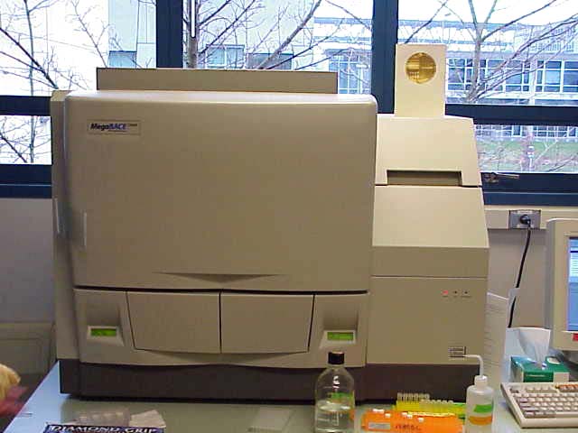
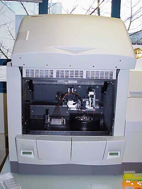


Figure 1. This is the Megabase Sequencer and Genotyper.
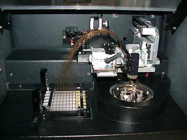
Figure 2. This what it looks like under the hood. The brown wire-like loops are the 96 individual capillary tubes. They are made of glass and coated with brown plastic. The samples are loaded from below this level on the left side and the DNA is electrophoresed towards the right. The matrix is injected into the tubes after each run and it travels from right to left when filling the tubes. See capillary electrophoresis explanation page.
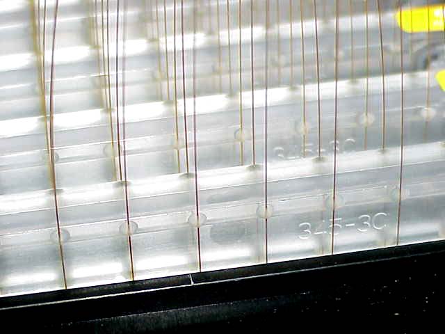
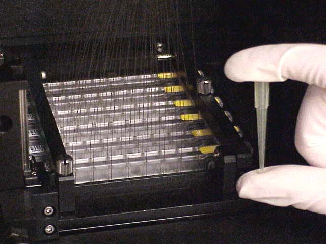
Figure 3. This is a close up of a few of the 96 capillary tubes. They continue down below this level and are submerged into the 96 well plate so DNA can enter the tubes. The photo on the right gives you an idea of the scale of the tubes which are 100 microns in diameter.
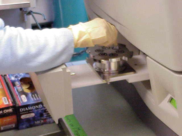
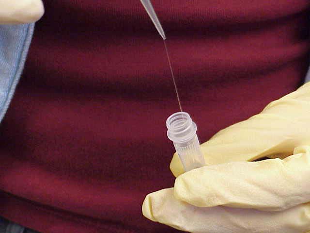
Figure 4. Heather is changing the resin tubes which are good for only a few days. The resin is a sticky polymer, as shown on the right.
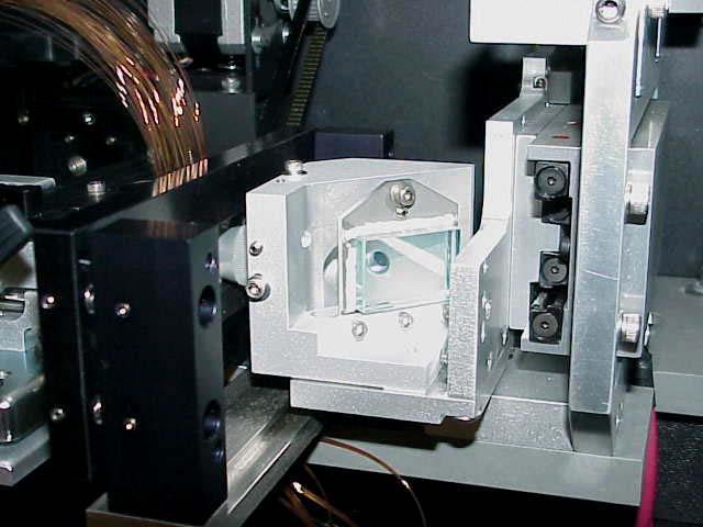
Figure 5. At the center of this photo is the scanner which passes a laser light through a short section of each capillary tube where there is no plastic coating. The laser excites the fluorophores and the emitted light (four colors, one for each base) is detected by a photomultiplier tube.
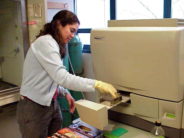
Figure 6. Heather is loading a 96 well plate into the chamber that will close and place the wells below the capillary tubes and later the plate will rise to submerge the open ends of the 96 tubes into the 96 DNA samples.
See the movie of the samples being loaded.
You'll be surprised how simple it is.
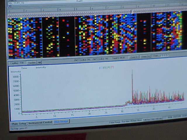
Figure 7. This image is from the computer screen showing the output of all 96 tubes. Using the mouse, you can take a closer look at the output from a single tube.
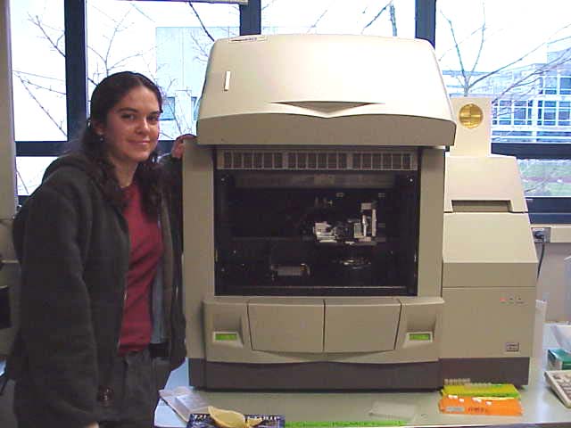
Figure 8. Even though much of this is automated, it still takes a well trained technician to keep it running smoothly each day. Heather is very attached to her Megabase, which always polite and informs her what to do next. :-)
© Copyright 2001 Department of Biology, Davidson College, Davidson, NC 28036
Send comments, questions, and suggestions to: macampbell@davidson.edu