This web page was produced as an assignment for an undergraduate course at Davidson College.
Prostaglandin Synthase (PGS) is an integral membrane protein located primarily in the endoplasmatic reticulum. Prostaglandins are involved in cell and tissue processes such as inflamation and platlet accumulation, threrefore, this protein is targetted by drugs like aspirin and ibuprofen. This page gives some general information about the structure and genetic sequence of the PGS proteins.

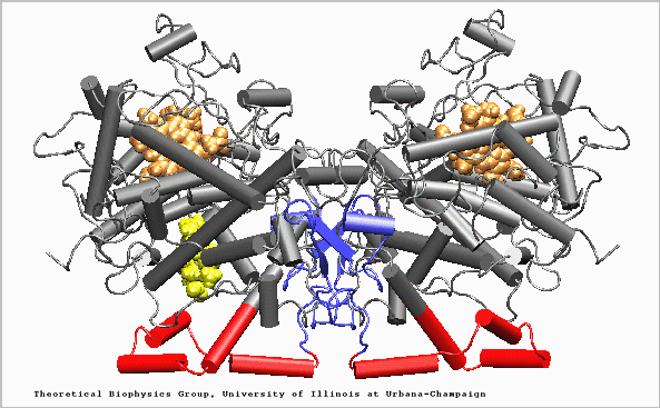
Figure 1. Structure of the Prostaglandin H2 synthase-1 homo-dimer: Epidermal Growth Factor-like domain (blue), amphipathic
membrane binding motif (red), catalytic globular domain (grey),
heme groups (brown), and arachidonic acid in
its binding site (yellow).
Through X-ray crystallography the structure of PGS can be determined. One example of PGS, prostaglandin H2 synthase-1 (PGHS-1), has three independent primary units: an epidermal growth factor domain, a membrane-binding motif, and an enzymatic domain (see Figure 1). The site critical for non-steroidal anti-inflammatory drug interactions is also a cyclo-oxygenase active site. This site is created by a long, hydrophobic channel.
View a 3-D, interactive Rasmol image of PGHS-1 here:
Sequence similarity of a certain protein among different organisms is a useful comparison for functionality comparisons as well as tracking evolution. The five genomic organisms listed below each contain genomic information on PGS in those organisms.
| Organism | Amino acid sequence | Nucleotide sequence |
| Homo sapiens | No |
gene |
| Mustela vison | Yes |
cDNA |
| Mus musculus | Yes |
mRNA |
| Gallus gallus | Yes |
mRNA |
| Bos taurus | Yes |
mRNA |
| Homo sapiens | Yes |
cDNA |
| Ovis sp. | Yes |
cDNA |
All genetic information searches were conducted on the National Center for Biotechnology Information webite.
Author: Aaron J.
Patton
Return to Aaron's Molecular Biology Switchboard
Return to the International House of Aaron
Return to Davidson's
Molecular Biology Homepage
(c)2000